- Tissue Microarrays (TMAs) Digital Library
- KS (HIV related)
- Lymphoma (HIV related)
- Lymphoma (HIV negative)
- Non-AIDS-defining Cancers (HIV related)
- Multsite Autopsies (HIV related NHL)
- Multsite Autopsies (HIV related KS)
- Multisite Autopsies (HIV related Lung Cancer)
- Antiretrovirals in Kaposi Sarcoma (KS-ARKS)
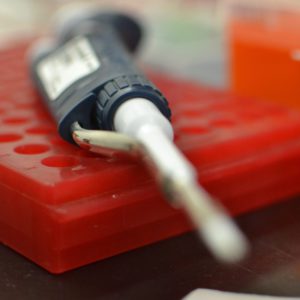
Investigators requesting access to view Tissue Microarray (TMA) images are required to complete the ACSR’s Virtual Biorepository Access Agreement. High level groupings of TMAs are listed in the menu to the left which link to a brief description of the individual TMA images available for viewing online.
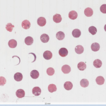
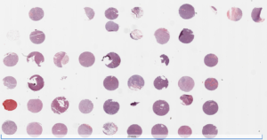
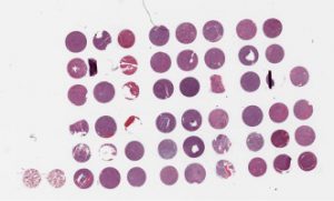
Tissue Microarray (TMA) sections are distributed with an accompanying diagram. The TMA diagram will show the relative placement of the patient tumor and orientation/control cores. The investigator will also receive a Quality Assessment summary for the materials received. This QA document will contain a pathologist’s review of the H&E for presence of core tissue and presence of tumor, as well as documentation of biological stains done to assess the tumor and nucleic acid integrity of each core. This QA may or may not have been done on a section near or adjacent to the sections you have received for your research. The percent of cores present is dependent upon the section number, the type of tissue and the method used to construct the TMA, therefore the total number of cores listed is an approximation based on the original number of cores embedded.
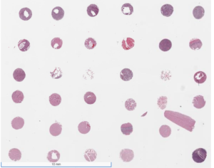
KS (HIV related) oral, visceral and skin from the U.S. and sub-Saharan Africa (SSA). Examples of TMA images are provided in the "Image" links below. Please go to our ACSR Digital Library for the complete collection of TMA images.
- KS TMA #11 Oral 2015: Approx. 19 tumor cores from 19 cases (Limited availability; please inquire) Image
- KS TMA #12 Oral 2015: Approx. 19 tumor cores from 19 cases (Digital images only; TMA not available) Image
- KS TMA #13 Visceral 2015: Approx. 28 tumor cores from 28 cases Image
- KS TMA #14 Visceral 2015: Approx. 28 tumor cores from 28 cases Image
- KS TMA #15 Skin 2015: Approx. 28 tumor cores from 28 cases (Limited availability; please inquire) Image
- KS TMA #16 Skin 2015: Approx. 28 tumor cores from 28 cases (Digital images only; TMA not available) Image
- SSA KS TMA #3 2010: Approx. 44 tumor cores from 28 cases Image
- SSA KS TMA #4 2010: Approx. 38 tumor cores from 23 cases Image
- SSA KS TMA #5 2010: Approx. 36 tumor cores from 24 cases Image
- SSA KS TMA #17 Oral 2016: Approx. 9 tumor cores from 9 cases Image
- SSA KS TMA #18 Visceral 2016: Approx. 9 tumor cores from 5 cases Image
- SSA KS TMA #19-1 Skin 2016: Approx. 28 tumor cores from 28 cases Image
- SSA KS TMA #19-2 Skin 2016: Approx. 27 tumor cores from 27 cases Image
- SSA KS TMA #19-3 Skin 2016: Approx. 9 tumor cores from 9 cases Image
- SSA KS TMA #20 Skin 2016: Approx. 32 tumor cores from 32 cases Image
- SSA KS TMA #21 Visceral 2016: Approx. 8 tumor cores from 4 cases Image
Lymphoma (HIV related) from the U.S. Examples of TMA images are provided in the "Image" links below. Please go to our ACSR Digital Library for the complete collection of TMA images.
- Misc. Lymphoma TMA 2010: Approx. 50 tumor cores from 31 cases Image
- DLBCL HIV+ TMA #4 2014: Approx. 10 tumor cores from 10 cases Image
- DLBCL HIV+ TMA #5 2014: Approx. 10 tumor cores from 10 cases Image
- DLBCL HIV+ TMA #8 2014: Approx. 54 tumor cores from 43 cases Image
- DLBCL HIV+ TMA #9 2014: Approx. 54 tumor cores from 45 cases Image
- Burkitt Lymphoma TMA #1 2017: Approx. 40 tumor cores from 26 cases Image
- Burkitt Lymphoma TMA #2 2017: Approx. 29 tumor cores from 19 cases Image
- Burkitt Lymphoma TMA #3 Bone Marrow 2017: Approx. 8 tumor cores from 6 cases Image
Lymphoma (HIV negative) from the U.S. Examples of TMA images are provided in the "Image" links below. Please go to our ACSR Digital Library for the complete collection of TMA images.
- PTLD TMA #1 2013: Approx. 11 tumor cores from 11 cases Image
- PTLD TMA #2 2013: Approx. 11 tumor cores from 11 cases Image
- Misc Lymphoma TMA #1 2014: Approx. 52 tumor cores from 26 cases Image
- Misc lymphoma TMA #2 2014: Approx. 52 cores from 25 cases Image
- DLBCL HIV negative TMA #6 2015: Approx. 38 cores from 38 cases Image
- DLBCL HIV negative TMA #7 2015: Approx. 38 tumor cores from 38 cases Image
Non-AIDS-defining Cancers (HIV related) from the U.S. Examples of TMA images are provided in the "Image" links below. Please go to our ACSR Digital Library for the complete collection of TMA images.
ANAL
-
- Anal Invasive TMA #1 2014: Approx. 12 tumor cores from 24 cases Image
- Anal Invasive TMA #2 2014: Approx. 10 tumor cores from 10 cases Image
- Anal In situ TMA #3 2014: Approx. 19 tumor cores from 19 cases: Limited number of pre-cut slides available, unable to make new fresh cuts Image
- Anal In situ TMA #4 2014: Approx. 7 tumor cores from 7 cases: Limited number of pre-cut slides available, unable to make new fresh cuts Image
- Anal Invasive TMA #1 2018: Approx. 16 tumor cores from 13 cases Image
- Anal Invasive TMA #2 2018: Approx. 12 tumor cores from 10 cases Image
- Anal In Situ TMA #3 2018: Approx. 8 tumor cores from 7 cases: Limited number of pre-cut slides available, unable to make new fresh cuts Image
- Anal Brazilian TMA #1 2021: Approx. 14 tumor cores from 13 cases Image
- Anal Brazilian TMA 2 2021: Approx. 14 tumor cores from 13 cases Image
- Anal Brazilian TMA 3 2021: Approx. 14 tumor cores from 14 cases Image
BREAST
-
- Breast TMA #1 2017: Approx. 10 tumor cores from 10 patients Image
HEAD AND NECK
-
- Head and Neck TMA #1 2018: Approx. 27 tumor cores from 25 cases Image
HODGKIN'S DISEASE
LUNG
PROSTATE
PRIMARY CENTRAL NERVOUS SYSTEM LYMPHOMA (PCNSL)
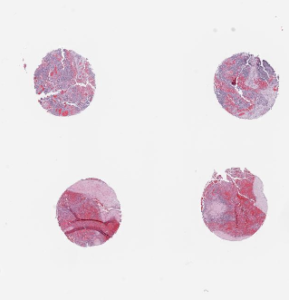
Multisite Autopsies (HIV related non-Hodgkin lymphoma) includes involved and uninvolved tissues. Examples of TMA images are provided in the "Image" links below. Please go to our ACSR Digital Library for the complete collection of TMA images.
- NHL TMA #9 2016: Approx. 7 tumor cores and 18 benign cores from 1 case (Limited availability; please inquire) Image
- NHL TMA #10 2016: Approx. 7 tumor cores and 18 benign cores from 1 case (Limited availability; please inquire) Image
- NHL TMA #11 2016: Approx. 16 tumor cores and 18 benign cores from 1 case (Limited availability; please inquire) Image
- NHL TMA #12 2016: Approx. 12 tumor cores and 22 benign cores from 1 case (Limited availability; please inquire) Image
- NHL TMA #13-1 2016: Approx. 20 tumor cores and 12 benign cores from 1 case (Limited availability; please inquire) Image
- NHL TMA #13-2 2016: Approx. 4 tumor cores and 17 benign cores from 1 case (Limited availability; please inquire) Image
- NHL TMA #14-1 2016: Approx. 18 tumor cores and 14 benign cores from 1 case (Limited availability; please inquire) Image
- NHL TMA #14-2 2016: Approx. 4 tumor cores and 17 benign cores from 1 case (Limited availability; please inquire) Image
Multsite Autopsies (HIV related Kaposi Sarcoma) includes involved and uninvolved tissues. Examples of TMA images are provided in the "Image" links below. Please go to our ACSR Digital Library for the complete collection of TMA images.
- KS TMA #1 2016: Approx. 4 tumor cores and 13 benign cores from 1 case (Limited availability; please inquire) Image
- KS TMA #2 2016: Approx. 3 tumor cores and 13 benign cores from 1 case (Limited availability; please inquire) Image
- KS TMA #3 2016: Approx. 4 tumor cores and 9 benign cores from 1 case (Limited availability; please inquire) Image
- KS TMA #4 2016: Approx. 8 tumor cores and 14 benign cores from 1 case (Limited availability; please inquire) Image
- KS TMA #5 2016: Approx. 4 tumor cores and 14 benign cores from 1 case (Limited availability; please inquire) Image
- KS TMA #6 2016: Approx. 4 tumor cores and 14 benign cores from 1 case (Limited availability; please inquire) Image
- KS TMA #7 2016: Approx. 3 tumor cores and 17 benign cores from 1 case (Limited availability; please inquire) Image
- KS TMA #8 2016: Approx. 3 tumor cores and 16 benign cores from 1 case (Limited availability; please inquire) Image
Multsite Autopsies (HIV related Lung Cancer) includes involved and uninvolved tissues. Examples of TMA images are provided in the "Image" links below. Please go to our ACSR Digital Library for the complete collection of TMA images.
- Autopsy Lung TMA #15 2019: Approx. 5 tumor cores and 10 benign cores from 1 case (Limited availability; please inquire) image
- Autopsy Lung TMA #16 2019: Approx. 7 tumor cores and 11 benign cores from 1 case (Limited availability; please inquire) image
- Autopsy Lung TMA #17 2019: Approx. 2 tumor cores and 15 benign cores from 1 case (Limited availability; please inquire) image
A randomized trial conducted from 2007 to 2011 to evaluate the efficacy of a protease inhibitor-based antiretroviral therapy regimen compared with a non-nucleoside reverse transcriptase inhibitor-based regimen to treat HIV-related histologically-confirmed KS in adults in Uganda. Data and biospecimens collected at multiple time points from a cohort of >200 participants. Examples of TMA images are provided in the "Image" links below. Please go to our ACSR Digital Library for the complete collection of TMA images.
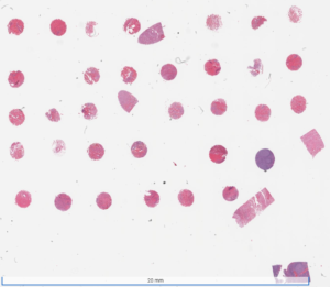
- ARKS TMA 1 2020: Approx. 37 tumor cores from 37 cases image
- ARKS TMA 2 2020: Approx. 36 tumor cores from 36 cases image
- ARKS TMA 3 2020: Approx. 35 tumor cores from 35 cases image
- ARKS TMA 4 2020: Approx. 36 tumor cores from 36 cases image